What type of scan do I need?

Do you need a scan at all?
The musculoskeletal system is made up of many types of tissue like bone, cartilage, ligaments, tendons, muscle and nerves as well as other tissues such as synovium and fascia that you may have heard of too. All of these structures are capable of causing pain and, as physiotherapists, it is our job to piece together all the information we get from the patient to understand what is causing the problem so we can then plan a route to recovery. A question we are commonly asked is “do I need a scan?”
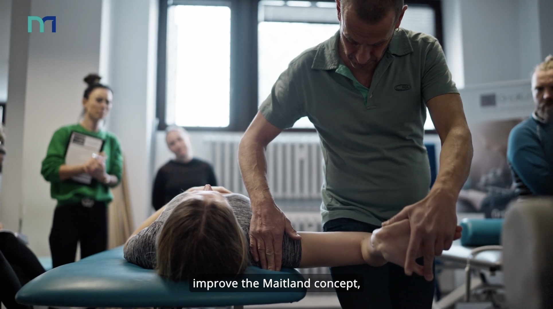
Before we start talking about scans, it’s important to understand that for every musculoskeletal complaint – whether it’s a spinal problem, frozen shoulder, tennis elbow or osteoarthritis of the knee or hip, the examination that the physio does provides the vast majority of the information needed to diagnose the problem and commence treatment. Whilst every clinician knows this, it can be tempting to resort to the high-tech scans and neglect taking a careful history of the problem and a comprehensive physical examination – particularly if this is what the patient wants. Our practice is built around the foundations laid out in the opening paragraph of the textbook Practice Principal Paul Hattam wrote about the subject:
“Clinical examination is the bedrock of diagnosis in musculoskeletal (MSK) medicine. It enables the clinician to both assemble information and to interpret the findings in order to assist in identifying the nature and stage of the patient’s disease or injury, determine the need for further investigations, provide a prognosis, guide treatment and measure outcome. Its big advantage is that it can be used at any stage in the patient’s management, and reproduction of the symptoms provides not only immediate feedback but also reassuring evidence, to both clinician and patient, that the source of the pain has been identified. Physical tests are quick and convenient to perform and, in the hands of a skilled practitioner, facilitate appropriate and cost-effective intervention”.
Paul Hattam, Handbook of Special Tests in Musculoskeletal Examination: An evidence-based guide for clinicians (2020)
What are the main types of imaging for MSK disorders?
Imaging (e.g. X-rays, MRI, CT and Ultrasound scans) form an important role in refining the diagnosis and planning the management. Different conditions need different types of scans – let’s summarise:
X-rays
X-rays can be the first port of call because they are readily available and comparatively cheap but are used much more cautiously now than they once were because of concern about safety and because there are better imaging options available now. They are most valuable for detecting abnormalities in bone such as fractures, tumours, infections and deformities. X-rays are also helpful in showing arthritic changes that can confirm rheumatoid or osteo arthritis. The problem with X-rays is that they don’t show any soft tissues (such as muscles, bursae, disc, ligaments, tendons or nerves) and these tissues are capable of being the primary cause of pain in many conditions.
Computed Tomography (CT)
CT uses X-rays to create an image and so the risks associated with ionising radiation applies here too. CT is brilliant for imaging bone where a very detailed profile is achieved and is particularly useful if MRI is not recommended or unavailable. The amount of time a person spends undergoing CT is much less than the MRI for safety reasons and it can be better tolerated as a result.
Magnetic Resonance Imaging (MRI)
MRI was discovered in the early 1970’s and was gradually introduced into medical practice in the 1980’s. It revolutionised medical diagnostics and enhanced our understanding of how disease and injury affects the musculoskeletal system. MRI is capable of producing very clear images of all MSK tissues including muscles, spinal discs, ligaments and tendons and is the ‘go to’ choice for things like spinal injuries, severe ligament ruptures at the knee and labral tears at the shoulder. The waiting times for MRI can be lengthy, but the information they provide can be very valuable.
Ultrasonography (USS)
Ultrasonography (or ultrasound scanning – USS) is an extremely useful diagnostic tool which is being used more and more frequently to identify inflammation in and around joints and tears or inflammation of tendons (e.g. rotator cuff injuries at the shoulder). It uses the same technology that has been employed in obstetric/maternity units for years and allows rapid and accurate evaluation of many MSK structures. In the hands of an experienced sonographer, USS has been shown to be equal to or better than MRI in its diagnostic accuracy for some tendon injuries. Another benefit is that the tissues can be scanned whilst they are moving – which after all – is exactly what they do! This can be fascinating for the patient too as they observe their body move in real time!
As an alternative to MRI & CT, ultrasonography is less expensive and, unlike CT, involves no exposure to radiation. Ultrasonography is also used as a guide when a needle needs to be put into a joint (for example, to inject drugs or to remove joint fluid).
Here at The Physios we offer ultrasound scanning ‘in house’. You can find out more about this service here.
So, do you still want a scan?
The Pro’s
There’s no doubt that a timely scan for some conditions can provide a great deal of useful information that will guide the patient’s management. It can also serve to reassure the patient that there is nothing seriously wrong.
The Con’s
It is well known that an abnormal scan doesn’t always point the clinician to the source of the problem. That is because tissues undergo a certain amount of ‘wear & tear’ over time which can make them look abnormal even though they are functioning well. This can lead the clinician to jump to the incorrect conclusion leading to mis-diagnosis and prolongation of the problem.
In conclusion
Imaging of the musculoskeletal system is not straight-forward and great care should be taken to ensure further tests are necessary and appropriate so that the patient’s recovery is accelerated and not hindered. But don’t worry about it – your physio will guide you on whether imaging is necessary for your condition.